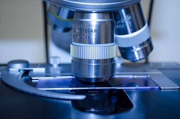
Dark-field microscopy
Vital blood examination in dark field according to Prof. Dr. G. Enderlein
The examination of a small drop of capillary blood under a dark-field microscope was developed by the zoologist and bacteriologist Prof Dr Günther Enderlein (1872-1968). The dark-field microscope can be used to observe microorganisms that live in the blood and were discovered and described by Prof Enderlein.
Here, the blood is observed and analysed under the microscope over a period of 24 hours - and even longer, until decomposition (with 100x and 400x and 1000x magnification).
The blood test is an almost indispensable part of diagnostics and subsequent therapy in practice. The procedure provides precise information about the composition of the blood, in particular about the condition of the white and red blood cells, the plasma and the microorganisms it contains. It provides information about the "inner environment" and the functionality of the blood cells as well as about the abundance and upward development of the smallest protein bodies (endobionts) present in every human being, from whose further development microorganisms and more highly developed structures such as bacteria, fungi or viruses can arise.
The presence of bacterial precursors, which are not yet disease-causing but put the body at risk of disease, can also be detected in the dark field examination. The dark field examination of the blood is therefore also a valuable preventive examination.
Dark field diagnostics is not only an excellent method for recognising diseases at a very early stage, it also offers therapy safety and an incorruptible therapy success control.
SANUM's isopathic medicines (fungal and bacterial preparations) and immunobiological agents profoundly change the endobiontic system of the symbionts by being able to reverse the pathological stages of upward development without having an antibiotic effect.
The selection of these remedies can be determined in the dark field.
PREPARATION: Fasting - appointments only possible in the morning! (for evening appointments - do not eat for 6 hours beforehand, drinking water is permitted) At the first appointment, a blood sample is taken from the fingertip or earlobe, for which a drop of blood is required. An immediate analysis is carried out, which the customer can observe on a large screen. A special experience! The blood is then observed, analysed and documented for 24 hours.
In a second appointment (no longer sober), the evaluation is discussed and a personalised treatment plan is handed over and the next steps are discussed in detail.
COST: € 150
Duration: 2x approx. 1 hour each
Book an appointment with Andrea Tillinger 0660 3453705


You are welcome to arrange a diagnostic appointment!
Ordination Klagenfurt:
Andrea Tillinger
Tel +43 (660) 3453705
andrea-tillinger.com


